Antibody-fluorochrome conjugates (called immunoconjugates) are extensively tested against normal and leukaemic cell targets using 2-, 3-, or 4-colour flow cytometry before being passed for general use in the various diagnostic or research applications.
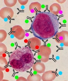
In the figure below, two distinct types of white blood cell - the smaller cells are red-blood cells - a granulocyte (lower left) and a myeloblast (upper right) have been exposed to four different types of fluorochrome-coupled antibodies. The fluorochromes are are coloured dyes, and are represented by the coloured circles attached to the antibodies (black structures). Green represents FITC, red is phycoerythrin (PE), purple is PE:Cy5 and blue is allophycocyanin (APC). The cells and antibody reagents are not drawn to scale, otherwise the cells would need to be over 1000 times larger, or about 15 metres diameter.
The granulocyte has both FITC- and PE-labelled antibodies attached, whereas the myeloblast has FITC- and PE:Cy5-labelled antibodies attached. Neither cell has been recognised by the APC-labelled antibodies.
In this example, assume the FITC-labelled antibody recognises a protein common to all myeloid cells, the PE-labelled antibody recognises 'mature' myeloid cells, PE:Cy5-labelled antibody recognises 'immature' myeloid cells, and the APC-labelled antibody recognises lymphocytes. Analysing a sample of these cells by flow cytometry would give two populations, one with green and red fluorescence only, the other with green and purple fluorescence only. From this we would conclude we had a mixed population of exclusively myeloid cells (since they were not recognised by the anti-lymphocyte reagent), one population was mature (granulocytes) and the other immature (myeloblasts). We would also be able to determine the relative frequency of each cell type in the sample.
Using these techniques, we can use many combinations of markers against different types of cell and stages of development to gain a comprehensive 'picture' of the different types of cells in any blood sample.
 The region responsible for recognising and binding to antigen targets is the antibody binding or Fab region, and contains the hypervariable regions of the two heavy- and light-chains (depicted in red in diagram opposite). The rest of the immunoglobulin molecule is relatively conserved in amino-acid structure (depicted in blue in diagram opposite), and is similar to other immunoglobulin molecules of the same class, and is known as the 'fragment-crystalline' or Fc region, and is responsible for cell-signalling after antibody has bound to its target.
![]()
Comments & feedback to: admin@hmds.org.uk