[ Home Page ]   [ Site Map ]

Chronic Lymphocytic Leukaemia


Clinical Features
Laboratory Diagnosis
Cytogenetics
Outcome and Therapy

CLINICAL FEATURES

Chronic lymphocytic leukaemia is the commonest of the lymphoproliferative disorders and is characterised by:

Top of Page


LABORATORY DIAGNOSIS

A) Morphology

[Reference 1]

Small cell morphology; click to enlarge (27K) Marrow infiltration by CLL cells; click to enlarge (40K)
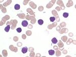
Small cells with thin rim of cytoplasm
Higher power magnification showing 'smear cell' (arrowed)
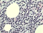
Bone marrow trephine biopsy showing extensive infiltration by CLL cells

B) Immunophenotype

[References 1,2]

Four-colour flow cytometric analysis of peripheral blood from a case of chronic lymphocytic leukaemia. The CLL cells co-express CD19 and CD5, but at a lower density than the corresponding 'normal' B-cells and T-cells. There is light chain restriction with almost all the CD19-positive CLL cells expressing sIg-kappa.

Top of Page


CYTOGENETICS

[References 7-12]

Atypical CLL: Trisomy 12 demonstrated by FISH technique
Atypical CLL cells showing trisomy 12, demonstrated by FISH (only visible in right-hand cells; out of focal plane in cell on left).
Top of Page


OUTCOME AND THERAPY

Top of Page


[ Home Page ] [ Site Map ]

Copyright © HMDS 1999-2005
Document last updated: Tuesday, 18 November 2003

Comments & feedback to: admin@hmds.org.uk
[URL: www.hmds.org.uk/cll.html]