[ Home Page ] [ Site Map ]

Mantle Cell Lymphoma


Clinical Features
Laboratory Diagnosis
Cytogenetics
Outcome and Therapy

CLINICAL FEATURES

Top of Page


LABORATORY DIAGNOSIS

A) Morphology

Replacement of nodal architecture, which may be diffuse or nodular.
A monomorphic population of small cells with irregular nuclei.
Nuclear expression of bcl-1 indicative of the t(11;14) translocation.

CD20-immunoperoxidase; click to enlarge (38K) Peripheral blood picture; click to enlarge (44K)
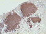
Intestinal MCL - CD20 immunoperoxidase staining of Peyer's patches.
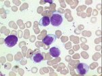
Involvement of peripheral blood and bone marrow is a very frequent finding in mantle cell lymphoma

B) Immunophenotype


CD5-PE vs CD19-Cy5; click to enlarge (7K) kappa-FITC vs lambda-PE; click to enlarge (6K) CD10-FITC vs CD19-Cy5; click to enlarge (5K)
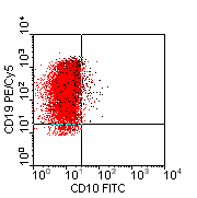
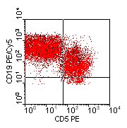
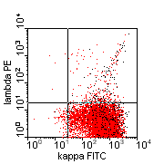
Three-colour flow cytometric analysis of a typical case of mantle cell lymphoma. There is strong co-expression of CD5 and CD19. CD23 is absent. The other main features are moderate to strong expression of sIg (IgD and IgM) with CD19 and CD20, and absence of CD10.

Top of Page


CYTOGENETICS

Cells with t(11;14) have one red, one green and one co-localised signal (arrows).


Immunoglobulin

bcl-1

Top of Page


OUTCOME AND THERAPY

Top of Page


[ Home Page ] [ Site Map ]

Copyright © HMDS 1999-2005
Document last updated: Tuesday, 18 November 2003

Comments & feedback to: admin@hmds.org.uk
[URL: www.hmds.org.uk/mcl.html]